
EEG - ElectroEncephaloGraphEEG is an acronym for
Electroencephalograph. This is a
recording ("graph") of electrical signals ("electro") from the brain
("encephalo"). They are made on chart paper that moves underneath pens
that are connected to galvanometers that read the electrical signals from
electrodes on the scalp. These electrodes do not send any electricity to
the person. They only receive electrical signals naturally generated by
the brain. Alpha waves are detected at the eight electrode positions indicated in the diagram. Usually, feedback is generated for the following four positions: O1, O2, C3, C4. O is for Occipital, C is for Central, F is for Frontal, and T is for Temporal. History of the Discovery of the EEGRichard Caton, a Liverpool physician and medical school lecturer, discovered electrical brain signals by probing directly on the surface of exposed brains of animals. He published his results in 1875. Caton used a reflecting galvanometer which was invented in 1858 by Lord Kelvin. Small changes in the position of a mirror attached to galvanometer coils produces a much larger movement of a reflected spot of light. This was a primitive form of amplification. Such instruments were capable of measuring micro-amperes. In 1887, Caton reported to the Ninth International Medical Congress in Washington, D.C. that when he interrupted light falling on an animal's eye, he detected negative variations in the electrical activity of the brain. It is ironic that he had to use a flame because electric light bulbs and electric power distribution were not yet widespread. He also discovered that electrical activity occurred in the opposite side of the brain from the eye. Most physiologists were unaware of Caton's work because they read scientific journals and he published in medical journals. Adolph Beck of Poland repeated and republished the work about 15 years after Caton in 1890 in a physiological journal. He went beyond Caton when he found that though a sensory stimulus such as a flash or a sound clap induced a response at a single point, there was a brain-wide interruption of the slow even pattern of waves in the brain. It was not until 1949 that the reticular activating system of the brain was discovered, which has an important role in controlling brain state. Fleischel von Marxow had made a similar discovery and placed the results in a sealed letter in a vault in 1883, a cowardly way of hedging scientific bets, as shown by the fact that he did not reveal it until after Beck's results were published. Marxow's attempt to claim credit was trumped when Caton wrote to the journal reminding them of his prior publication. A Russian scientist, Vasili Yakovlevich Danilevsky had made similar experiments in 1876 and published them in his doctoral thesis in 1877 (two years after Caton). Between Danilevsky and Beck, Nikolai Y. Wedensky had used a telephone to listen to electrical waves in the brains of cats and dogs. In 1912, Vladimir V. Pravdich-Neminsky published photographic recordings of brain waves in dogs.
Dr. Hans Berger, an Austrian psychiatrist was the first to record electroencephalographs from humans. After gaining his doctorate at the University of Jena in 1897, Berger became aware of Richard Caton's work. His experiments with animals were inconclusive by 1910, but after World War I he decided to look for the EEG in the human brain. Though he worked with primitive instruments such as string galvanometers he was a shy and secretive man so he did not seek help. In the early years of the 1920s Berger obtained his first results in subjects who had skulls with gaps under the skin where bone was missing. He made recordings on moving photographic paper with a wavy spot of light. This was how Berger found the regular waves at about 10 cycles per second that he named the Alpha waves because they were the first waveforms he isolated in the human EEG. Berger published a paper in 1929 based on the research he had done five years earlier with his son Klaus as a subject. He made 73 recordings, which became the first published EEGs of humans. Berger found that the best recordings were made with an electrode on the occiput (lower rear of the skull) and another on the forehead (which functions as a reference). During the five years, the cautious Berger made recordings from himself and many other subjects. He wanted to eliminate the possibility that blood circulation might be creating harmonics in the recordings, so he made many recordings with simultaneous electrocardiograms and blood pressure from the head. Berger also eliminated the possibility that the waves originated in the skin by doing some experiments with electrodes just below the skin with special insulation. In 1929, Berger wrote "The electroencephalogram represents a continuous curve with continuous oscillations in which ... one can distinguish larger first order waves with an average duration of 90 milliseconds and smaller second order waves of an average duration of 35 milliseconds [Beta waves]. The larger deflections measure at most 150 to 200 microvolts...." "We see in the electroencephalogram a concomitant phenomenon of the continuous nerve processes which take place in the brain, exactly as the electrocardiogram represents a concomitant phenomenon of the contractions of the individual segments of the heart." A year later Berger has accumulated 1,133 recordings from 76 subjects. In a paper he published that year Berger first named the Alpha and Beta waves and began to use the initials EEG for ElectroEncephaloGram. Berger reported that the amplitude of the Beta waves was smaller than Alpha, despite the fact that he was able to demonstrate that Beta waves were related to mental concentration and to startle reactions. In 1931 Berger found that alpha waves diminished during sleep, during general anaesthesia, and during cocaine stimulation. He reported that the Alpha wave frequency was decreased in patients with high intracranial pressure resulting from injuries. Berger also found, significantly, that epileptic patients had large amplitude waves. He also wrote that Alzheimer's disease and multiple sclerosis altered EEGs. On the other hand Berger was disappointed to find that many illnesses had little or no obvious effect on the recorded EEG. These included schizophrenia, melancholia, manic-depression (bipolar illness), mental retardation, and aphasia (speech loss). Modern research by Dr. Hardt and others has shown by using a wider time perspective that integrated amplitude neurofeedback enhancement of alpha and other brain waves has useful effects for some of these types of sufferers. The Carl Zeiss Foundation took note of Berger's growing discoveries and gave him electronic amplifiers and a specially made oscillograph, along with an assistant. With this sensitive equipment he repeated and replicated his earlier work. Berger found that an epileptic patient had nearly flat waves after a seizure but alpha waves slowly increased as consciousness was regained. He recorded EEGs from young children and infants. Berger found that brain waves began to appear only at about two months of age. This corresponds to the time when the brain neurons become sheathed in myelin. For several years, Berger's work was ignored by scientists because many thought that overall brain wave recordings would just be confused roars and then because the alpha waves were "dull" regular waves. In 1934 Edgar Adrian and B.H.C. Matthews of the Cambridge Physiological Laboratory confirmed Berger's research and published their results in the journal Brain, where they referenced Berger's work. They used copper gauze electrodes wrapped in saline soaked lint. They wanted to call the Alpha waves the Berger rhythm, but Hans Berger was modest and rejected it. Berger soon became world famous, except in Germany. There Hitler's regime was oppressing his University of Jena. Thus he was greatly surprised when in 1937 at an international congress in Paris he found himself celebrated by researchers from around the world. However on September 30, 1938 he was forced by Nazi officials to retire the next day. After a series of further tragedies, Berger committed suicide in 1941. He was twice considered for the Nobel Prize, but the Nazis prevented it from being awarded and accepted. By contrast, in England two EEGs labs before the war had grown to 50 by the end because of the usefulness in diagnosing brain injuries. MEG - MagnetoEncephaloGramMEG is a new technology that holds great promise because it can detect brain waves deep in the brain. However, if the brain's electrical field is weak when measured at the scalp, the brain's magnetic field is weaker still, especially when compared to the earth's magnetic field and motor magnets. It requires large superconducting magnets, and today that requires liquid gases to get cold enough. Contact BI by e-mail or phone at 650-941-9999. Copyright 2000-2002 Biocybernaut Institute Mountain View, California |


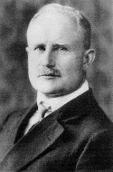 Dr. Hans Berger